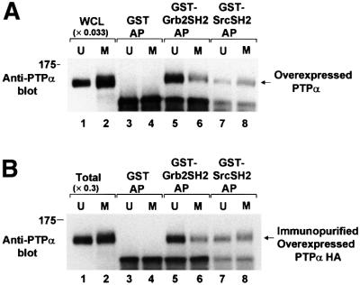
Fig. 5. In vitro binding of unsynchronized and mitotic PTPα to the Grb2 and Src SH2 domains. (A) Lysates from unsynchronized (U, odd lanes) or mitotic (M, even lanes) PTPα-HA overexpressor cells were affinity-precipitated by incubation with GST (lanes 3 and 4), a GST–Grb2 SH2 domain fusion protein (lanes 5 and 6) or a GST–Src SH2 domain fusion protein (lanes 7 and 8) bound to Sepharose beads. The washed beads were then analyzed by 9% SDS–PAGE and anti-PTPα immunoblotting. For comparison, lanes 1 and 2 (WCL) contained 0.033 times the amount of complete whole cell lysate. (B) PTPα-HA was immunopurified from unsynchronized or mitotic overexpressor cells and affinity-precipitated by GST or the GST–SH2 domain fusion proteins used above, analyzed by 9% SDS–PAGE and immunoblotted with anti-PTPα antibody (lanes 3–8). For comparison, lanes 1 and 2 (Total) contain 0.3 times the amount of immunopurified PTPα used in the affinity precipitations. The positions of molecular weight standards are indicated in kDa.

An official website of the United States government
Here's how you know
Official websites use .gov
A
.gov website belongs to an official
government organization in the United States.
Secure .gov websites use HTTPS
A lock (
) or https:// means you've safely
connected to the .gov website. Share sensitive
information only on official, secure websites.
