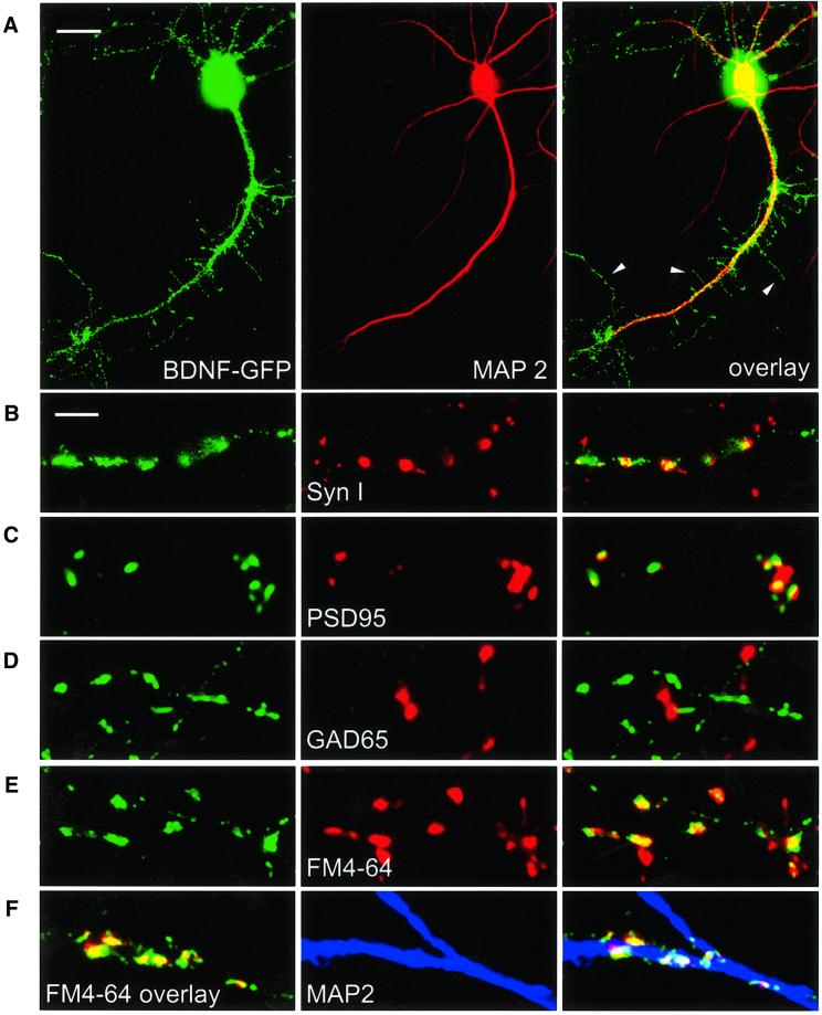
Fig. 1. Synaptic targeting of BDNF–GFP. Immunofluorescent detection of the dendritic marker protein MAP2 (A), the general presynaptic marker protein Synapsin I (B), the marker protein PSD95 for glutamatergic synapses (C) and the marker protein GAD65 for GABAergic synapses (D), respectively. (E) Identification of presynaptic terminals in living neurons using activity-dependent labelling with FM 4-64. In (A–E), each panel shows dendrites of a BDNF–GFP-expressing hippocampal neuron (left), the fluorescence image for the respective marker (middle) and the overlay of both (right). (F) Left: live staining of FM 4-64-labelled synaptic terminals (red) of a BDNF–GFP (green)-expressing neuron shows synaptic localization of BDNF–GFP (yellow). Middle: posthoc labelling of dendrites using MAP2 antibody (blue). The overlay on the right shows co-localization of the three signals in white. Scale bars: 10 µm (A), 4 µm (B–F). The arrowheads in (A) depict dendritic filopodia.

An official website of the United States government
Here's how you know
Official websites use .gov
A
.gov website belongs to an official
government organization in the United States.
Secure .gov websites use HTTPS
A lock (
) or https:// means you've safely
connected to the .gov website. Share sensitive
information only on official, secure websites.
