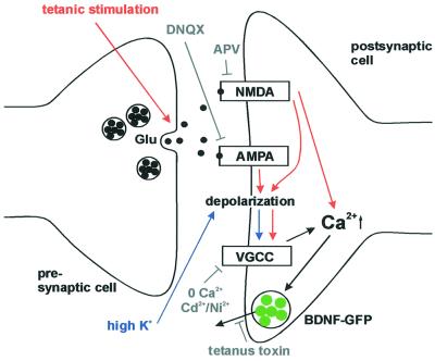
Fig. 6. Synaptic secretion of BDNF–GFP by different routes of postsynaptic depolarization. Tetanic presynaptic stimulation leads, via intermediate repetitive glutamate release, to postsynaptic depolarization and Ca2+ influx (red route). This depolarization can be blocked by ionotropic glutamate receptor antagonists (see Figure 5). In contrast, high K+ solution directly depolarizes the postsynaptic membrane (blue route), activates VGCCs, and can thus bypass intermediate glutamate release and activation of postsynaptic glutamate receptors (see Figure 3). The observed inhibition of BDNF release by blocking Ca2+ influx (0 Ca2+; Ni2+/Cd2+) and the independence of BDNF release from glutamate receptors (both during stimulation with high K+) demonstrate the direct dependence of BDNF secretion on postsynaptic Ca2+ influx. Likewise, since K+-induced postsynaptic depolarization is independent of glutamate release, the inhibition of high K+-induced BDNF secretion by tetanus toxin indicates that BDNF secretion depends on the fusion of secretory vesicles.

An official website of the United States government
Here's how you know
Official websites use .gov
A
.gov website belongs to an official
government organization in the United States.
Secure .gov websites use HTTPS
A lock (
) or https:// means you've safely
connected to the .gov website. Share sensitive
information only on official, secure websites.
