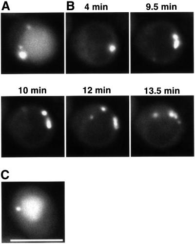
Fig. 9. Time-lapse microscopy of apg1ts cells expressing GFP–Aut7p. Δapg1 cells (NNY20) carrying the apg1ts and GFP–Aut7p plasmids were used. (A) Cells treated with rapamycin for 3 h at 30°C. (B) Time-lapse images following temperature decrease. Cells were treated with rapamycin for 4 h at 37°C before decreasing the temperature to 30°C. (C) Cell incubated for 2 h at 30°C after temperature decrease. Bar: 5 µm.

An official website of the United States government
Here's how you know
Official websites use .gov
A
.gov website belongs to an official
government organization in the United States.
Secure .gov websites use HTTPS
A lock (
) or https:// means you've safely
connected to the .gov website. Share sensitive
information only on official, secure websites.
