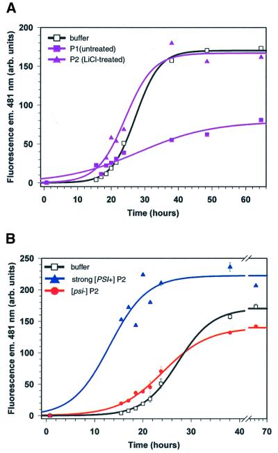
Fig. 2. Sucrose cushion pellet proteins from [PSI+] but not [psi–] lysates increased the rate of NM conversion. (A) Kinetics of NM fiber formation with or without proteins pelleted from the ΔNMSup35 strain. NM fiber formation was monitored by fluorescence emission of thioflavin-T (ThT) at 481 nm. Two fractionation methods were used, preparation 1 (untreated, see Materials and methods) or 2 (LiCl treated). The black curve (open squares) represents the progress of NM conversion in the presence of buffer diluted 25-fold; the purple curve with filled triangles represents [psi–] ΔNMSup35 pelleted P2 proteins diluted 25-fold and the purple curve with filled sqaures represents [psi–] strain ΔNMSup35 pelleted P1 proteins diluted 100-fold. (B) Effect of different salt-treated P2 pellets on the kinetics of NM conversion. Conversion was monitored by fluorescence emission of ThT at 481 nm. Sucrose cushion P2 pellets or buffer were diluted 25-fold. Curves are drawn to indicate the likely data trends and are not correlated sigmoidal fits.

An official website of the United States government
Here's how you know
Official websites use .gov
A
.gov website belongs to an official
government organization in the United States.
Secure .gov websites use HTTPS
A lock (
) or https:// means you've safely
connected to the .gov website. Share sensitive
information only on official, secure websites.
