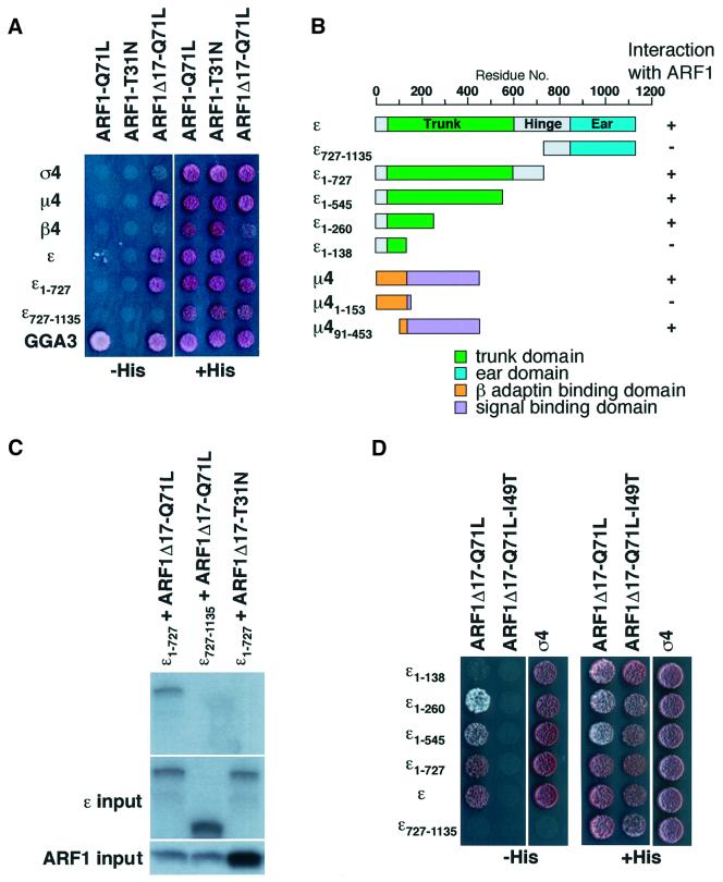
Fig. 5. Interaction analyses between ARF1 and AP-4 subunits and truncation analyses of the ε–ARF1 interaction. (A and D) HF7C yeast strain was transfected with constructs expressing the indicated proteins and co-transformants were spotted on plates with (right) or without (left) histidine. Interaction of proteins was assessed by growth on the plate lacking histidine. (B) Bar diagram summarizing the results depicted in (A), (D) and Figure 6A. (C) In vitro transcribed/translated 35S-labeled ε and ARF-myc constructs were mixed, incubated, and ARF-myc was immunoprecipitated using an anti-myc antibody. Top, the co-precipitation of ε1–727 was detected by autoradiography. Middle and bottom, labeled ε and ARF1 constructs, respectively, which were used as input.

An official website of the United States government
Here's how you know
Official websites use .gov
A
.gov website belongs to an official
government organization in the United States.
Secure .gov websites use HTTPS
A lock (
) or https:// means you've safely
connected to the .gov website. Share sensitive
information only on official, secure websites.
