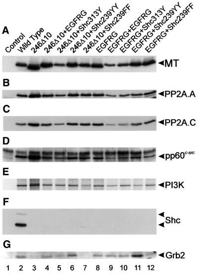
Fig. 2. Cellular polypeptides associated with MT mutants. Stable cell lines expressing each of the MT mutants shown in Figure 1 were lysed, the MT immunoprecipitated with PAb762, separated by SDS–PAGE and western blotted. MT-associated proteins were detected in duplicate blots by the use of specific antibodies. The probe used to detect each polypeptide is shown on the right together with the migration position. For detection of the 85 kDa subunit of PI3K, MT immunoprecipitates were incubated with [γ-33P]ATP, separated by SDS–PAGE and autoradiographed. The mutant used is indicated above each lane. Only the 52 and 66 kDa forms of ShcA were detected. The faint band migrating slightly faster than the 52 kDa form of ShcA is a non-specific reaction caused by the heavy chain of the immunoprecipitating PAb762. The experiments shown are representative of five different experiments performed with two different sets of cell lines.

An official website of the United States government
Here's how you know
Official websites use .gov
A
.gov website belongs to an official
government organization in the United States.
Secure .gov websites use HTTPS
A lock (
) or https:// means you've safely
connected to the .gov website. Share sensitive
information only on official, secure websites.
