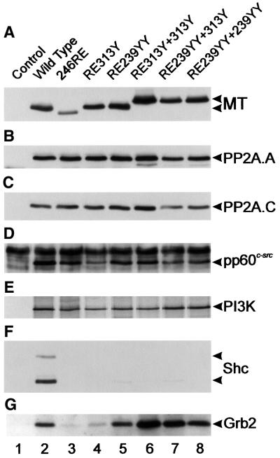
Fig. 4. Polypeptides associated with the MT mutants shown in Figure 3. Cell lines expressing the MT mutants shown in Figure 3 were lysed, the MT immunoprecipitated, blotted and probed with the antibodies shown on the right. Association with the PI3K 85 kDa subunit was detected by an in vitro kinase reaction. The mutant used is indicated above each lane. The experiment was repeated six times on two different sets of cell lines with similar results.

An official website of the United States government
Here's how you know
Official websites use .gov
A
.gov website belongs to an official
government organization in the United States.
Secure .gov websites use HTTPS
A lock (
) or https:// means you've safely
connected to the .gov website. Share sensitive
information only on official, secure websites.
