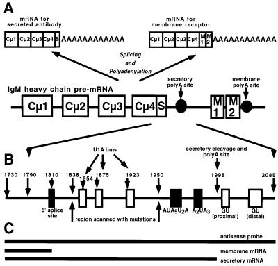
Fig. 1. Schematic model of the immunoglobulin secretory poly(A) site. (A) The genetic organization of the IgM heavy chain and its alternative processing to a secretory or a membrane form of mRNA. (B) The location of the secretory poly(A) site and relative location of the 5′ splice site, the U1A-binding motifs, the hexanucleotide sequence and the downstream GU-rich regions. Numbers indicate the positions of the ends of the RNA substrates and plasmid inserts referred to in the text. (C) The length and position of the antisense probe used for RNase protection assays and the protected fragments for the µ secretory and µ membrane mRNA, respectively, referred to in Figures 6 and 7.

An official website of the United States government
Here's how you know
Official websites use .gov
A
.gov website belongs to an official
government organization in the United States.
Secure .gov websites use HTTPS
A lock (
) or https:// means you've safely
connected to the .gov website. Share sensitive
information only on official, secure websites.
