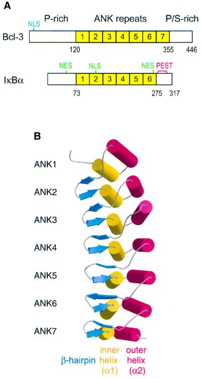
Fig. 1. Structure of Bcl-3. (A) Domain organization of Bcl-3 and IκBα. The basic nuclear localization sequence (NLS) of Bcl-3 is indicated in blue, while the leucine-rich NLS and NES of IκBα are in green. The fragment of Bcl-3 that was crystallized spans residues 119–359 and encompasses the entire ARD. (B) Ribbon diagram of the Bcl-3 ARD. The molecule curves towards the α1 helices, which together with the β-hairpins forms the presumed binding surface for p50 and p52 homodimers. This figure and Figures 3B and C, 5 and 6 were made with Bobscript (Esnouf, 1999) and Raster3D (Merritt and Bacon, 1997).

An official website of the United States government
Here's how you know
Official websites use .gov
A
.gov website belongs to an official
government organization in the United States.
Secure .gov websites use HTTPS
A lock (
) or https:// means you've safely
connected to the .gov website. Share sensitive
information only on official, secure websites.
