
Figure 4.
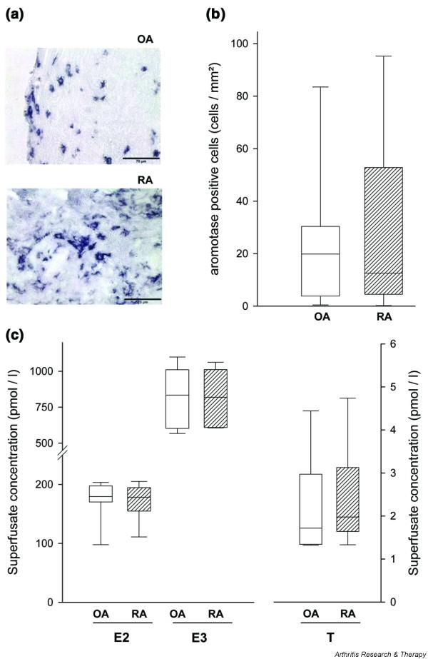
Aromatase expression in synovial tissue and endogenous steroid hormone release from superfused synovium. (a) Immunohistochemistry of aromatase in one OA and one RA patient. Using the respective control antibody revealed no staining of positive cells (not shown). Magnification: 400×. (b) Density of aromatase-positive cells in OA (open bars, n = 20) and RA patients (hatched bars, n = 16). (c) Spontaneously released E2, E3, and free testosterone from standardized superfused pieces of synovial tissue of OA (open bars, n = 20) and RA patients (hatched bars, n = 18). (b,c) Values are given as box blots with the 5th, 25th, 50th (median), 75th, and 95th percentile when applicable. OA, osteoarthritis; RA, rheumatoid arthritis. Other abbreviations are as given in the legend to Fig. 1.