
Figure 2.
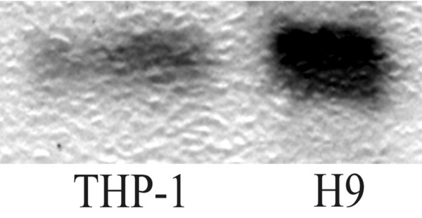
Western blot analysis of anti-TIMP-2 antibodies. Lysates of THP-1 (a human monocytic cell line) and H9 (a human T-cell lymphoma) were separated in 18% Tris-glycine gel, transferred into a polyvinylidene fluoride membrane, and blotted with immunoglobulin G (IgG) fractions from a patient with rheumatoid arthritis having high levels of anti-TIMP-2 antibodies detected by ELISA. The IgG fraction visualized a band of molecular weight 22 kDa, corresponding to TIMP-2. TIMP, tissue inhibitor of metalloproteinases.