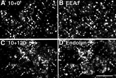
Fig. 5. Apically internalized endolyn is delivered to lysosomes. Polarized MDCK cells expressing endolyn were incubated with apically added anti-endolyn mAb for 10 min, washed and incubated without antibody for 0 min (A and B) or 120 min (C and D). After fixation, cells were double-labeled with rabbit antibodies to EEA-1 (B) or endolyn (D), followed by incubation with Cy3 anti-mouse and FITC-anti-rabbit antibodies, and analyzed by confocal microscopy. Arrows point to structures that contain both endocytosed mAb and the respective marker; bar, 10 µm.

An official website of the United States government
Here's how you know
Official websites use .gov
A
.gov website belongs to an official
government organization in the United States.
Secure .gov websites use HTTPS
A lock (
) or https:// means you've safely
connected to the .gov website. Share sensitive
information only on official, secure websites.
