Figure 2.

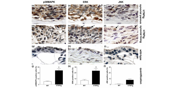
Expression of MAPK in synovial lining and pannus cells of hTNFtg mice and wild-type mice. The phosphorylated forms of (a,d,g) p38 mitogen-activated protein kinase (MAPK)α, (b,e,h) extracellular signal-regulated kinase (ERK), and (c,f,i) c-Jun amino-terminal kinase (JNK) were stained in both the synovial pannus (panels a–c) and the synovial lining layer (panels d–f) of human tumour necrosis factor transgenic (hTNFtg) mice as well as in wild-type mice (panels g–i). p38MAPKα and ERK were abundantly activated in hTNFtg mice but not in wild-type mice (brown staining, black arrows). In contrast, JNK was activated far less frequently and only in a few cells within synovial pannus as well as the synovial lining in hTNFtg mice. In wild-type mice, activation of MAPKs was generally low. Original magnification 1000×. Quantitative analysis showed a significantly higher expression of (j) p38MAPKα and (k) ERK in hTNFtg mice compared with wild-type mice, but no significant difference in (l) JNK activation. Data are expressed as mean ± standard error of the mean. *P < 0.05. WT, wild-type.
