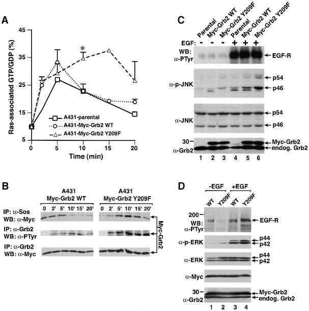
Fig. 8. Mutation of Grb2 Tyr209 potentiates and prolongs EGF signaling pathways. (A) Ras activation. A431 parental cells and populations stably expressing wild-type Myc-Grb2 and Myc-Grb2 Y209F were starved, labeled with [32P]orthophosphate and stimulated with EGF (200 ng/ml) for 0, 2, 5, 10, 15 or 20 min, and Ras-associated guanine nucleotides analyzed as described in Materials and methods. Bars indicate positive SE for the A431-Myc-Grb2 WT and A431-Myc-Grb2 Y209F cells; the 15 min time point was only done once. The difference in Ras GTP levels at 10 min between wild-type Grb2 and Grb2 Y209F cells (asterisk) is statistically significant (P ≤0.05, unpaired t-test). (B) Kinetic analysis of Grb2 tyrosine phosphorylation and Sos association from the A431-Myc-Grb2 WT- and A431-Myc-Grb2 Y209F-expressing cells in (A). Cells were starved and stimulated with EGF for the indicated times and lysates analyzed by immunoprecipitation with anti-Sos (top panel) or anti-Grb2 (middle and bottom panels) antibodies and blotting with anti-Myc (top and bottom panels) or anti-PTyr (middle panel) antibodies. (C) JNK activation. Parental (lanes 1 and 4), Myc-Grb2 WT- (lane 2 and 5) and Myc-Grb2 Y209F-expressing (lanes 3 and 6) A431 cells were starved (lanes 1–3) and stimulated with EGF (200 ng/ml, lanes 4–6), and lysates analyzed by western blot using anti-PTyr antibody for detecting EGF-stimulated phosphorylation of EGF receptor (top panel), anti-phospho-JNK antibody (second panel from the top), anti-JNK antibody (third panel from the top) or anti-Grb2 antibody (bottom panel). (D) MAPK/ERK activation. 293T cells transfected with EGF receptor and either Myc-tagged wild-type Grb2 (lanes 1 and 3) or Myc-tagged Grb2 Y209F (lanes 2 and 4) were starved (lanes 1 and 2) or stimulated with EGF for 10 min (200 ng/ml, lanes 3 and 4), and lysates were analyzed by western blot using anti-PTyr antibody for detection of phosphorylated EGF receptor (top panel), anti-phospho-ERK antibody (second panel from the top), anti-ERK antibody (third panel from the top), anti-Myc antibody (fourth panel from the top) and anti-Grb2 antibody (bottom panel).

An official website of the United States government
Here's how you know
Official websites use .gov
A
.gov website belongs to an official
government organization in the United States.
Secure .gov websites use HTTPS
A lock (
) or https:// means you've safely
connected to the .gov website. Share sensitive
information only on official, secure websites.
