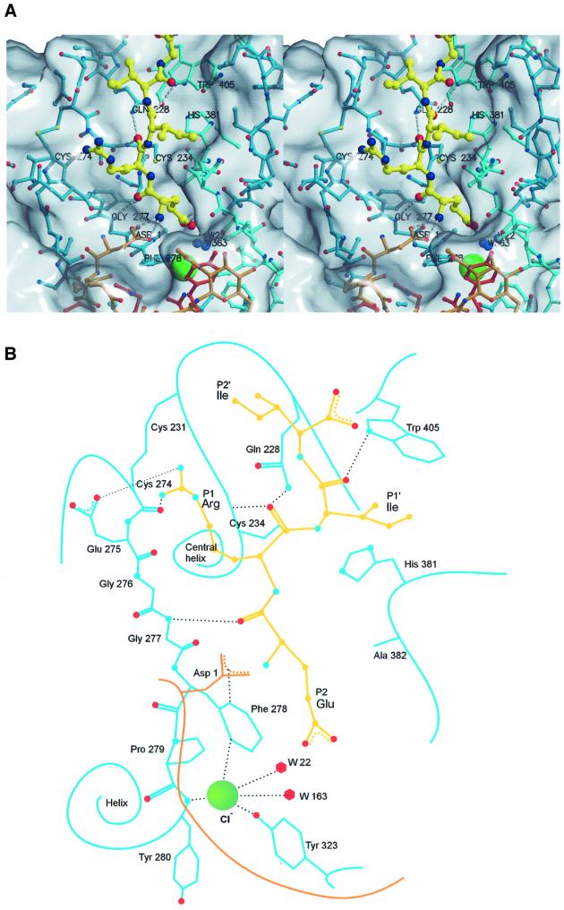
Fig. 3. Active site cleft of DPPI with a bound model of the N-terminal sequence ERIIGG from the biological substrate, granzyme A. (A) Stereo view: covalent bonds of papain-like domains and the exclusion domain are shown in the colors used in Figure 1C. Covalent bonds of the substrate model are shown as yellow sticks. Corresponding carbon atoms are shown as balls using the covalent bond color scheme. The chloride ion is shown as a large green sphere. Oxygen, nitrogen and sulfur atoms are shown as red, blue and yellow spheres, respectively. The residues relevant for substrate binding are marked and hydrogen bonds are shown as white broken lines. The molecular surface was generated with GRASP (Nicholls et al., 1991); the figure was prepared in MAIN (Turk, 1992) and rendered with RENDER (Merritt and Bacon, 1997). (B) Schematic presentation. The color codes are the same as in (A).

An official website of the United States government
Here's how you know
Official websites use .gov
A
.gov website belongs to an official
government organization in the United States.
Secure .gov websites use HTTPS
A lock (
) or https:// means you've safely
connected to the .gov website. Share sensitive
information only on official, secure websites.
