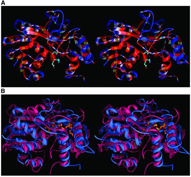
Fig. 3. (A) A ribbon stereodiagram showing the overall fold of NmrA with secondary structure labelled. The conserved residues among the NmrA and NMR1s (Figure 1) are coloured red whilst non-conserved residues are coloured blue. The side chains of three conserved charged residues on helix α6 are shown. (B) Stereodiagram comparing the overall structure of NmrA (blue) and UDP-galactose 4-epimerase (red). The bound NAD and UDP-glucose are shown as ball and stick representations.

An official website of the United States government
Here's how you know
Official websites use .gov
A
.gov website belongs to an official
government organization in the United States.
Secure .gov websites use HTTPS
A lock (
) or https:// means you've safely
connected to the .gov website. Share sensitive
information only on official, secure websites.
