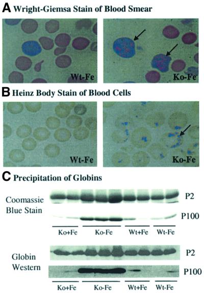
Fig. 5. Precipitation of globins in RBCs of HRI–/– mice in iron deficiency. (A) Wright–Giemsa-stained peripheral blood smears. Peripheral blood smears were prepared from HRI +/+ and –/– mice maintained on an iron-deficient diet for 33 days. (B) Heinz body staining. Blood from mice at day 92 after receiving a low iron diet was collected and stained with crystal violet for the presence of Heinz bodies. (C) Precipitation of globins in the blood cells of HRI–/– mice. Precipitated proteins from the blood of Wt and HRI–/– mice at day 67 after receiving a low iron diet were collected by centrifugation and separated by 15% SDS–PAGE. Proteins were analyzed by Coomassie Blue staining (upper two panels) and by western blot analysis using anti-mouse hemoglobin antibody (lower two panels). P2, pellets of 2000 g centrifugation; P100, pellets of 100 000 g centrifugation.

An official website of the United States government
Here's how you know
Official websites use .gov
A
.gov website belongs to an official
government organization in the United States.
Secure .gov websites use HTTPS
A lock (
) or https:// means you've safely
connected to the .gov website. Share sensitive
information only on official, secure websites.
