Figure 6.
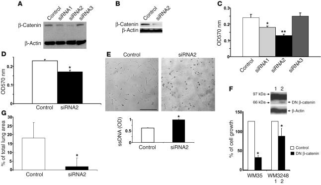
Suppressing β-catenin reverses Notch1-induced tumor growth and metastasis. (A and B) β-catenin was examined by immunoblotting in WM35–NIC-GFP cells expressing β-cat–siRNA1, β-cat–siRNA2, β-cat–siRNA3 and control vector (A) and in WM3248–NIC-GFP cells expressing β-cat–siRNA2 (B). β-cat–si–RNA2 efficiently suppressed expression of β-catenin. β-actin was used as loading control. (C and D) MTT assay shows a decreased growth rate induced by β-cat–siRNA1 (*P < 0.01, Student’s t test) and β-cat–siRNA2 (**P < 0.001, Student’s t test) in WM35–NIC-GFP cells and β-cat–siRNA2 in WM3248-NIC-GFP cells when compared with control. Results are mean ± SD from triplicates of 3 independent experiments. (E) Induction of cell apoptosis by β-cat–siRNA2 in WM35–NIC-GFP cells. Round shape indicates cell death. Scale bar: 20 μm. Cell apoptosis was quantitatively measured by ELISA. Results are mean ± SD from triplicates of 3 independent experiments. Similar results were observed in WM3248–NIC-GFP cells. (F) 3[H]-Thymidine incorporation assay shows a decreased growth rate of WM3248 cells transfected with DN β-catenin compared with the control. Results are mean ± SD from 3 independent experiments. Inner panel shows expression of DN β-catenin protein. β-actin was used as loading control. (G) In vivo lung colony–formation assay. β-cat–siRNA-WM3248-NIC-GFP or H1UG-WM3248-NIC-GFP cells were injected intravenously into mice. After 12 weeks, lung samples were harvested and analyzed. Tumor colonies formed on the lung surface were macroscopically counted under a dissection microscope. Data are mean ± SD.

