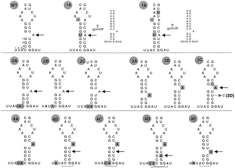
Fig. 2. Schematic secondary structure of D6 mutants. Mutations to the abbreviated D6 sequence are shown in bold and highlighted in gray. The name of each variant is indicated to the upper left. The branch site of each mutant is indicated with an arrow. For the WT variant, the 7 nt downstream from D5 are shown. For mutants 1A and 1B, the two possible equilibrium conformations of D6 are indicated. For mutants 4B–E, brackets indicate additional base pairs that may serve to extend the D6 stem. RNA 3D is shown as a mutant of 3C, from which it is derived, with an arrow indicating the site of uridine to cytosine mutation.

An official website of the United States government
Here's how you know
Official websites use .gov
A
.gov website belongs to an official
government organization in the United States.
Secure .gov websites use HTTPS
A lock (
) or https:// means you've safely
connected to the .gov website. Share sensitive
information only on official, secure websites.
