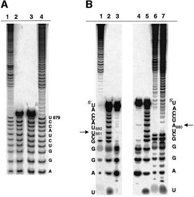
Fig. 4. Analysis of branch-site choice for intron revertants. (A) The branch points for mutants 1A (lane 4) and 1B (lane 1) were mapped by limited alkaline hydrolysis of branched fragments labeled at their 5′-termini. These were subjected to electrophoresis next to alkaline hydrolysis ladders of an oligonucleotide marker corresponding to the 17 nt immediately upstream of the WT branch-point adenosine (lanes 2 and 3). For the mapping procedure employed (Figure 3), the last band before the gap indicates the nucleotide 5′ of the selected branch point (A880, in each of these cases). (B) Partial alkaline hydrolysis of 3′-labeled branched fragments. For the mapping procedure employed (Figure 3), the band directly beneath the gap will correspond to a position 2 nts downstream from the branch point. Thus, the gap beginning at position C882 (lanes 6 and 7) corresponds to a WT branch-site choice at position A880 for mutants 1A (lane 6) and the prA–G (lane 7; Chu et al., 1998). Partial alkaline hydrolysis of these fragments is shown next to corresponding T1 (lane 4) and hydroxide (lane 5) ladders on an oligonucleotide marker corresponding to the terminal 13 nt of the intron. Partial alkaline hydrolysis of a cryptic branching mutant (RNA 4B, lane 1) is shown next to T1 (lane 3) and hydroxide (lane 2) ladders, and corresponds to branching at position U881 (arrow).

An official website of the United States government
Here's how you know
Official websites use .gov
A
.gov website belongs to an official
government organization in the United States.
Secure .gov websites use HTTPS
A lock (
) or https:// means you've safely
connected to the .gov website. Share sensitive
information only on official, secure websites.
