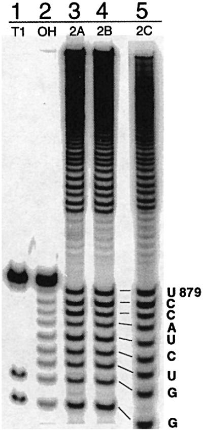
Fig. 5. Mapping the branch points of linker mutants 2A–C. Partial alkaline hydrolysis of branched fragments labeled at the 5′-end indicates that, like the WT intron, these mutants all branch at position A880 (lanes 3–5). The T1 digest (T1) and alkaline hydrolysis (OH) ladders on the 13 nt oligonucleotide serve as markers.
