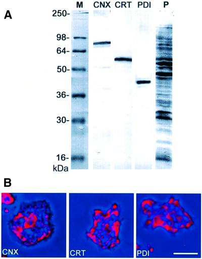
Fig. 2. Immunolabeling of endogenous calreticulin and calnexin in Dictyostelium cells. (A) Specificity of mAbs used as ER markers, assayed in western blots of total cellular proteins. CNX, calnexin labeled with mAb 270-390-2; CRT, calreticulin labeled with mAb 252-234-2; PDI, protein disulfide isomerase labeled with mAb 221-135-1. The last lane (P) shows a parallel lane stained for proteins with Ponceau S. (B) Immunofluorescence labeling of fixed cells using the antibodies shown in (A). Immunofluorescence in red is superimposed on phase-contrast images in dark blue. All three antibodies label the reticulate structure of the ER. The perinuclear layer in the center of the cell is most prominently labeled with the calnexin antibody. Bar in (B), 10 µm.

An official website of the United States government
Here's how you know
Official websites use .gov
A
.gov website belongs to an official
government organization in the United States.
Secure .gov websites use HTTPS
A lock (
) or https:// means you've safely
connected to the .gov website. Share sensitive
information only on official, secure websites.
