. 2005 Feb 15;113(6):729–734. doi: 10.1289/ehp.7791

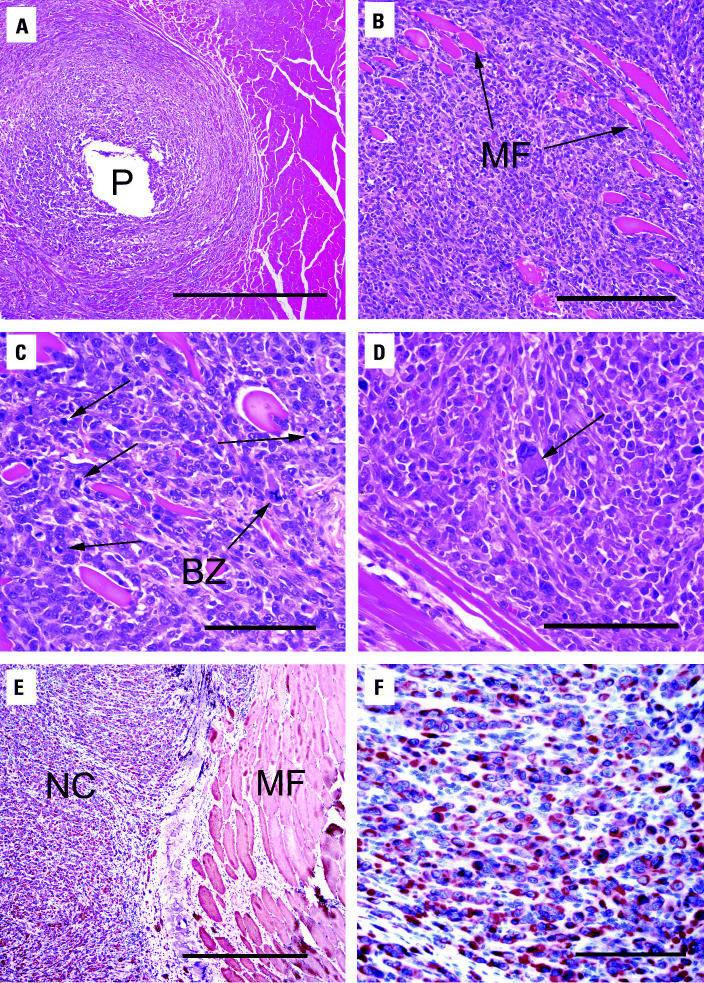
Figure 3. Histopathologic examination of leg tumor surrounding WA pellet. (A) H&E-stained section of leg tumor from F344 rat showing WA pellet hole (P); bar = 500 μm. (B) H&E-stained tumor section showing neoplastic infiltration of preexisting muscle fibers (MF); bar = 200 μm. (C) H&E-stained tumor section showing neoplastic cells with numerous mitoses (arrows) and bizarre mitotic figures (BZ); bar = 100 μm. (D) H&E-stained tumor section showing pleomorphic cell (arrow); bar = 100 μm. (E) Desmin staining of leg tumor showing neoplastic cells (NC) and muscle fibers (MF); bar = 500 μm. (F) Desmin staining of neoplastic cells; bar = 50 μm.

Figure 3