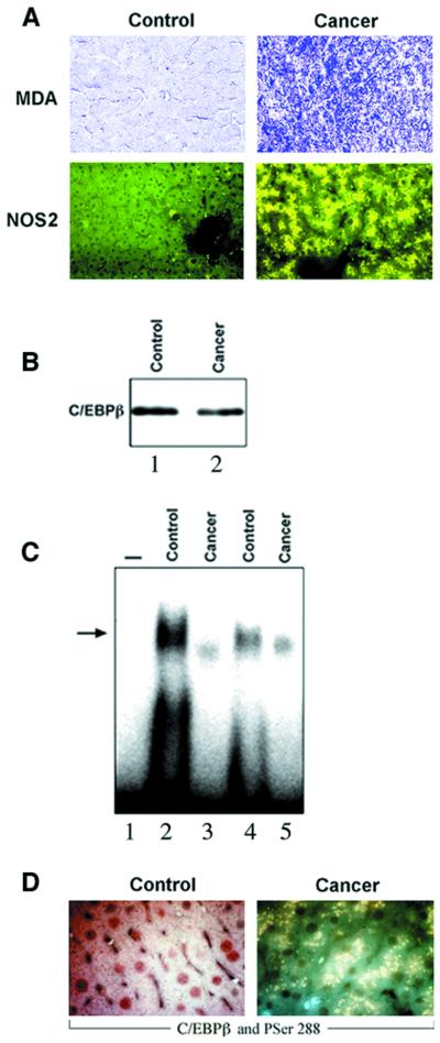
Fig. 7. Nuclear export of C/EBPβ-Ser288 in the liver of patients with cancer-cachexia. (A) Representative immunohistochemistry for MDA–protein adducts, NOS2, C/EBPβ and C/EBPβ-PSer288 of control individuals (control) and cancer-cachexia patients (cachexia) was performed as described in Figures 1 and 4. (B) A representative C/EBPβ protein immunoblot in C/EBPβ immunoprecipitates from liver protein lysates (250 µg) of control (lane 1) and cancer-cachexia (lane 2) subjects was performed as described in Materials and methods. (C) Mobility shift analysis of liver nuclear extracts (5 µg of protein) and the 32P-labeled D-site of the albumin enhancer/promoter (1 ng) was performed as described in Figure 2. The position of the bound DNA is indicated by an arrow. Samples shown are: control (lanes 2 and 4); cancer-cachexia (lanes 3 and 5). On lane 1, the probe was processed without nuclear extracts. (D) Representative immunohistochemistry using antibodies for C/EBPβ (in red) and C/EBPβ-PSer288 (in green), simultaneously. In control liver, C/EBPβ was localized in the nucleus and C/EBPβ-PSer288 was undetectable. In cancer-cachexia liver, C/EBPβ-PSer288 was detected in the cytoplasm in yellow due to the superimposition of C/EBPβ (red) and C/EBPβ-PSer288 (green).

An official website of the United States government
Here's how you know
Official websites use .gov
A
.gov website belongs to an official
government organization in the United States.
Secure .gov websites use HTTPS
A lock (
) or https:// means you've safely
connected to the .gov website. Share sensitive
information only on official, secure websites.
