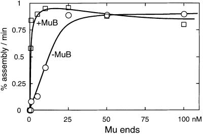
Fig. 5. MuB stimulates transpososome assembly at low Mu end concentrations. The assembly reactions were carried out as described in Materials and methods, except that the concentration of Mu end DNA was varied from 0.5 to 100 nM. The concentrations of MuA and MuB–target complex were optimized for each Mu end concentration. The assembly time course was analyzed as described for Figure 3, except that the labeled L-end that was incorporated into all transpososomes was counted and the percentage assembly rate was plotted against the Mu end concentration.

An official website of the United States government
Here's how you know
Official websites use .gov
A
.gov website belongs to an official
government organization in the United States.
Secure .gov websites use HTTPS
A lock (
) or https:// means you've safely
connected to the .gov website. Share sensitive
information only on official, secure websites.
