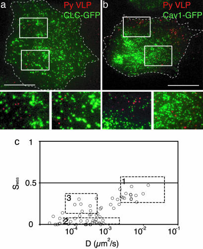
Fig. 4.
Confinement of VLPs on the cell surface does not require caveolae or clathrin-coated pits. TIRF images of AF568 VLPs bound to the bottom surface of live 3T6 cells expressing clathrin light chain–GFP (a) or caveolin-1–GFP (b). (Scale bars, 10 μm.) For each construct, a representative merged, dual-color image of a whole cell (Upper) and close-ups (insets in Upper shown in Lower) are shown. (c) D/SMSS plot of VLP trajectories from particles bound to lung fibroblasts obtained from caveolin-1 knockout mice.

