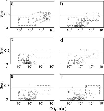
Fig. 5.
Effect of perturbations of cellular actin and cholesterol on VLP motion. D/SMSS plots show VLP mobility when bound to cells in the presence of 0.2 μM latrunculin A added 15 min before the VLPs (a), 0.25 μM jasplakinolide added in the same way (b), 10 mM MCD added 1 h before the VLPs (c), 10 mM MCD–cholesterol complex added for 2 h to cells pretreated with MCD as in c (d), 0.2 μM latrunculin A for 15 min in cells pretreated as in c (e), and 0.2 mM genistein for 1 h added before the VLPs (f).

