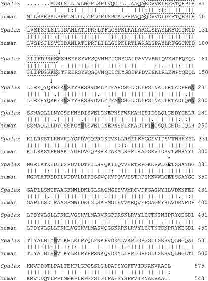
Fig. 2.
Comparison of Spalax and human heparanase amino acid sequences. Vertical lines denote conserved amino acids, and double or single dots mark similar amino acids (Wisconsin Package, Version 103). The putative two catalytic Glu residues, the proton donor and the nucleophile, are marked in bold with * above. The potential N-glycosylation sites are shaded. The 8-kDa subunit is marked with a dotted box. The cleavage sites generating the mature enzyme are marked by arrows, and amino acids between the two arrows denote the linker sequence. The sequence boxed with a continuous line denotes the amino acids lacking in splice 7 of the Spalax heparanase.

