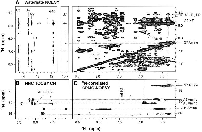
Fig. 1. Unusual NOEs of the AGAA hairpin, 5′-GGUUC[AGAA]GAACC. (A) Portions of the NOESY spectrum of the AGAA hairpin recorded at 293 K on a 2 mM unlabeled sample in H2O. Note the cross-peak between the imino proton (10.7 p.p.m.) and the amino proton (6.4 p.p.m.) resonances. The NOE cross-peaks between the G7 amino proton and the A6 ribose and H8 protons are indicated on the spectral region on the right. (B) Portion of the spectrum of an HNC-TOCSY-CH experiment recorded at 293 K on a 1.0 mM 13C,15N-labeled sample. The amino nitrogen resonances of the adenines are correlated with both the H2 and H8 resonances within the same base. (C) Portion of the 15N-correlated CPMG-NOESY spectrum at 293 K on a 1.0 mM 13C,15N-labeled sample. The intense cross-peaks are between intraresidue amino proton and amino nitrogen resonances.

An official website of the United States government
Here's how you know
Official websites use .gov
A
.gov website belongs to an official
government organization in the United States.
Secure .gov websites use HTTPS
A lock (
) or https:// means you've safely
connected to the .gov website. Share sensitive
information only on official, secure websites.
