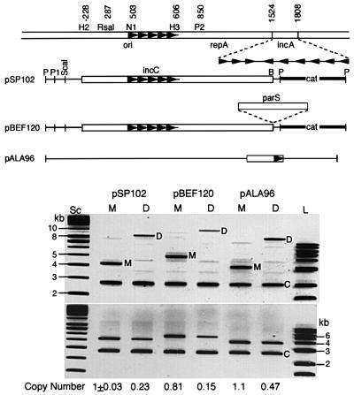
Fig. 1. Plasmid copy number in cells carrying either a plasmid monomer (M) or an isogenic plasmid dimer (D). Top: schematic map of plasmids. Open bars represent P1 DNA, thin lines pBR322 DNA, and thick lines a drug marker DNA. Coordinates in the top line are from Abeles et al. (1984). The enzyme names have been abbreviated in some cases as follows: HincII (H2), NruI (N1), HindIII (H3), PvuII (P2), PstI (P), PvuI (P1) and BamHI (B). Arrowheads represent iterons. The three genetic elements that comprise the P1 plasmid replicon are ori (387–611), initiator gene repA (664–1521) and the control locus, incA (1524–1808). pSP102 is a miniP1 plasmid. pBEF120 is identical to pSP102 except for the presence of parS. pALA96 is derived from pBR322 (Chattoraj et al., 1984). Bottom: lanes Sc and L represent supercoiled and linear DNAs that served as size markers in kb units. Plasmid DNA was isolated from cells carrying the two (M and D) forms. Before DNA isolation, a control culture containing pNEB193 (C) was added to each of the experimental cultures to account for loss during DNA isolation. The upper and lower panels are identical except that the DNAs were digested with HindIII in the lower panel for ease of measuring total plasmid DNA. The band intensities were normalized with respect to the pSP102 monomer band and adjusted further for plasmid size to determine relative copy numbers.

An official website of the United States government
Here's how you know
Official websites use .gov
A
.gov website belongs to an official
government organization in the United States.
Secure .gov websites use HTTPS
A lock (
) or https:// means you've safely
connected to the .gov website. Share sensitive
information only on official, secure websites.
