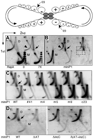
Fig. 6. Topological assay of physical interactions between miniP1 iterons in dimeric plasmids. Top: schematic showing simultaneous binding of a tetrameric protein to two diametrically opposite sites in a homodimer that blocks diffusion of + and – supercoils (Sc), generated by transcription (semicircular arrows), across the sites. Relaxation specifically of –Sc by a topoisomerase (ω) could result in accumulation of +Sc in the dimer. Protein binding to a single site cannot block Sc diffusion, hence no +Sc can accumulate (adapted from Wu and Liu, 1992). Topoisomers of plasmid M and D are separated by two-dimensional agarose gel electrophoresis. In the first dimension, plasmids with more Sc, + or –, migrate faster. In the second dimension, run in the presence of chloroquine, plasmids with +Sc run ahead of those with –Sc. The leading and trailing edges of the plasmid topoisomer distribution contain plasmids with most +Sc (arrow) and most –Sc (arrowhead), respectively. (A) Distribution of a pBR322-derived plasmid (pAO; Wu and Liu, 1992) to which the incC iterons have been added. The 0 and 7× RepA were supplied from compatible plasmids pST52 and pALA198, respectively (Park et al., 1998) (1× is physiological RepA). Plasmids with +Sc are seen only when RepA was supplied. (B) Topoisomer distribution of a miniP1 plasmid, pSP102. The dashed boxes show the region used to quantify the +Sc/–Sc ratio. The intensity of the lower two (empty) boxes was used as background and subtracted from the corresponding upper boxes before calculating the ratio. Plasmids with +Sc predominate in the D but not in the M population. (C) Relative levels of +Sc in pSP102 and its five high-copy derivatives (plasmids are the same as in Figure 3). From the comparison of band intensities of plasmids with +Sc and –Sc, from the two edges of the topoisomer distribution (arrow and arrowhead, respectively), it is apparent that there are relatively fewer plasmids with +Sc in mutants than in the wild-type (WT). (D) Participation of iterons in the generation of +Sc in plasmid dimers. One of the two origins is defective in mutant dimers (plasmids are the same as in Figure 4). Plasmids with +Sc are reduced particularly when the incC iterons are deleted [ΔincC and Δ(AT+incC), last two lanes]. Comparison of ΔincC and Δ(AT+incC) plasmids also shows that the AT-rich region contributes somewhat to the accumulation of +Sc.

An official website of the United States government
Here's how you know
Official websites use .gov
A
.gov website belongs to an official
government organization in the United States.
Secure .gov websites use HTTPS
A lock (
) or https:// means you've safely
connected to the .gov website. Share sensitive
information only on official, secure websites.
