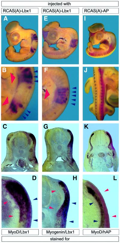
Fig. 1. Forced expression of Lbx1 stimulates myogenesis in the paraxial segmented mesoderm in vivo. HH25 chicken embryos were subjected to whole-mount in situ hybridization after injection of RCAS(A)–Lbx1 (A–H) and RCAS(A)–hAP (I–L) into somites I–IV or VI–X of HH10 embryos and 3 days of incubation. Depending on the injection procedure, which involved several injections per embryo, either two separate sets (A and B), a single block of somites (E and F) or most somites of one half (I and J) were infected. Two-color whole-mount in situ hybridization of MyoD (dark purple staining in A–D) and Lbx1 (red staining in A–H) and of myogenin (dark purple staining in E–H) and Lbx1. Embryos in I–L were hybridized with a MyoD antisense probe (dark purple staining) and stained for hAP activity (red staining). Whole-mount preparations (A, B, E, F, I and J) and vibratome sections (C, D, G, H, K and L) are shown. Forced expression of Lbx1 (indicated by bold red arrowheads) resulted in a strong up-regulation of MyoD (A–D) and myogenin (E–H) in somites (blue arrowheads in B, D, F and H), while the expression of hAP did not result in an up-regulation of MyoD (I–L) and other myogenic markers. A shorter staining time in RCAS(A)–Lbx1-injected embryos allowed only the detection of stimulated MyoD and myogenin expression, while uninjected halves showed only a very weak MyoD (C) and myogenin (G) signal.

An official website of the United States government
Here's how you know
Official websites use .gov
A
.gov website belongs to an official
government organization in the United States.
Secure .gov websites use HTTPS
A lock (
) or https:// means you've safely
connected to the .gov website. Share sensitive
information only on official, secure websites.
