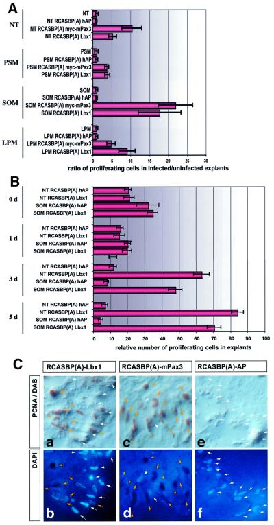
Fig. 5. Forced expression of Lbx1 and Pax3 strongly stimulates cell proliferation in embryonic mesoderm and NT explants. (A) Tissue fragments derived from NT (columns 1–4), PSM (columns 5–8), SOM (columns 9–12) and LPM (columns 13–16) were infected with RCAS–Lbx1, RCAS–Pax3 and RCAS–hAP viruses. Proliferating cells were identified by staining for PCNA antigen. Values on the x-axis indicate x-fold induction of the number of proliferating cells in infected versus uninfected explants. (B) Tissue fragments derived from SOM or NT were infected with either RCAS–hAP or RCAS–Lbx1 and analyzed for the presence of proliferating cells at different time points after explantation. Stimulation of proliferation by Lbx1 became apparent 3 days after infection and exceeded the relatively high proliferation rate in cultures without Lbx1 at day 0. Values on the x-axis indicate the number of proliferating cells relative to the absolute number of cells in explants. (C) Representative example for Lbx1- and Pax3-induced cell proliferation. SOM explants were immunostained with a PCNA antibody (a, c and e) 5 days after infection with RCAS–Lbx1 (a and b), RCAS–Pax3 (c and d) and RCAS–hAP (e and f) viruses. Nuclear DNA was visualized by DAPI staining (b, d and f). Yellow arrows mark PCNA-positive cells; white arrows mark non-proliferating cells. In explants infected with RCAS–hAP, very few proliferating cells were seen, and in RCAS–Lbx1 and RCAS–Pax3 infected explants numerous PCNA-positive, proliferating cells were detectable.

An official website of the United States government
Here's how you know
Official websites use .gov
A
.gov website belongs to an official
government organization in the United States.
Secure .gov websites use HTTPS
A lock (
) or https:// means you've safely
connected to the .gov website. Share sensitive
information only on official, secure websites.
