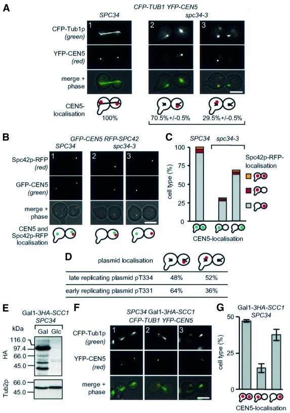
Fig. 6. Sister CEN5 DNAs of spc34-3 cells are associated preferentially with the old SPB. (A) SPC34 and spc34-3 cells with CFP-TUB1 YFP-CEN5 were arrested with α-factor to mark the mother cell. Washed cells were then shifted to 37°C for 3 h. CFP–Tub1p and YFP–CEN5 were analysed by fluorescence microscopy. The percentage of anaphase cells with YFP–CEN5 (red dot) at both or only one of the two spindle poles was determined in two independent experiments (n >100). The black lines in the drawn cells symbolize the MTs. The mating projection of the mother cell body is shown as an extension. (B) Cells of GFP-CEN5 RFP-SPC42 in which the old SPB is marked by the fluorescent RFP signal were grown as described in Materials and methods. The Spc42p–RFP and the GFP–CEN5 signals were detected by fluorescence microscopy. The red dot in the cartoons indicates the old SPB and the green dots the GFP–CEN5 signals. The mating projection marking the mother cell body is indicated as an extension. (C) Quantification of (B); n >100. Symbols are as in (B). (D) Preferential binding of sister chromatids to the old SPB is dependent on the time of centromere replication. Cells of spc34-3 with the late and early replicating plasmids pT334 and pT331 were analysed as in (A). Cartoon as in (A). (E) Gal1-3HA-SCC1 SPC34 cells carrying CFP-TUB1 YFP-CEN5 were grown and analysed as described in Figure 7A. (F) Cells of (E) were treated and analysed as in Figure 7B. (G) Quantification of (F). Shown is the average of two independent experiments; n >100. Cartoons are as in (A). Bars: 5 µm.

An official website of the United States government
Here's how you know
Official websites use .gov
A
.gov website belongs to an official
government organization in the United States.
Secure .gov websites use HTTPS
A lock (
) or https:// means you've safely
connected to the .gov website. Share sensitive
information only on official, secure websites.
