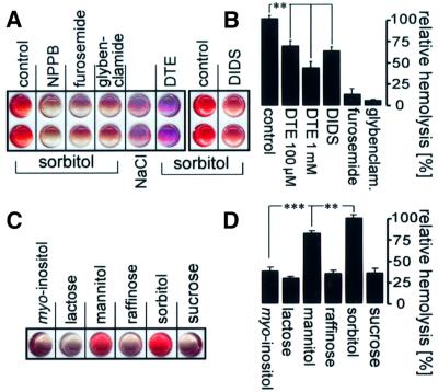
Fig. 4. Hemolysis of infected cells in isosmotic sorbitol solution. (A and B) Hemolysis was sensitive to reduction, NPPB, furosemide, glybenclamide and DIDS. Enriched trophozoite-infected erythrocytes were suspended in isosmotic sorbitol or, for control, in NaCl and incubated in the presence and absence of DIDS, NPPB, furosemide, glybenclamide (each 100 µM) and DTE (100 µM and 1 mM), respectively. Incubation was stopped by centrifugation, and hemolysis was indicated by hemoglobin in the supernatant. In (A) are the scanned images of supernatants from two individual experiments performed in duplicate. (B) Mean hemolysis in isosmotic sorbitol in the absence (control) and presence of DTE (100 µM and 1 mM, respectively), DIDS, furosemide and glybenclamide (each 100 µM), respectively. The hemoglobin concentration of the supernatants was determined photometrically and data were expressed as a percentage of that fraction of total hemolysis that could be inhibited by NPPB (n = 3–16; **P ≤0.01). (C and D) Substrate dependence of the infection-induced isosmotic hemolysis. In (C) are the imaged supernatants (individual experiment) and in (D) the mean relative hemolysis of cells incubated in different isosmotic carbohydrate solutions as indicated (n = 8–9; **P ≤0.01; ***P ≤0.001).

An official website of the United States government
Here's how you know
Official websites use .gov
A
.gov website belongs to an official
government organization in the United States.
Secure .gov websites use HTTPS
A lock (
) or https:// means you've safely
connected to the .gov website. Share sensitive
information only on official, secure websites.
