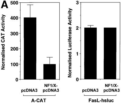
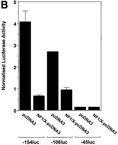
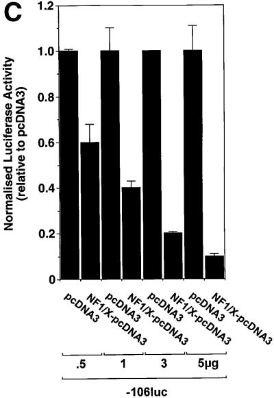
Fig. 3. Repression of PDGF-A promoter activity by NF1/X. (A) WKY12-22 cells were transiently co-transfected with 2 µg of A-CAT or 2 µg of a luciferase construct driven by 1.2 kb of the Fas ligand promoter, along with either 3 µg of NF1/X–pcDNA3 or pcDNA3. Twenty-four hours after transfection, the cells were lysed and assessed for CAT or luciferase activity. CAT activity was normalized to the concentration of protein in the lysate. (B) Serial 5′ deletion and transient transfection analysis using PDGF-A promoter constructs and NF1/X. COS-7 cells were transfected with the indicated PDGF-A promoter–luciferase constructs together with 3 µg of either NF1/X-pcDNA3 or pCDNA3. Twenty-four hours after transfection, the cells were lysed and assessed for luciferase activity. (C) Overexpression of NF1/X represses PDGF-A chain transcription in a dose-dependent manner. COS-7 cells were transiently transfected with 2 µg of –106luc and the indicated amounts of NF1/X or pcDNA3. Twenty-four hours post-transfection, the cells were lysed and assessed for luciferase activity. In experiments involving luciferase reporters, the cells were also transfected with 2 µg of pRL–TK to normalize for transfection efficiency. Firefly luciferase activity was normalized to Renilla. Identical results were obtained in WKY12-22 cells.

An official website of the United States government
Here's how you know
Official websites use .gov
A
.gov website belongs to an official
government organization in the United States.
Secure .gov websites use HTTPS
A lock (
) or https:// means you've safely
connected to the .gov website. Share sensitive
information only on official, secure websites.


