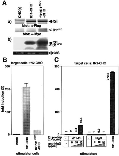
Fig. 6. Involvement of multimerization of Delta1 in the N2 activation. (A) Generation of fD1-CHO cells expressing Myc-tagged D1ΔICD (fD1/D1ΔICD-CHO). (a) Expression of Myc-tagged D1ΔICD and Flag-tagged fD1 proteins in fD1/D1ΔICD-CHO cells were examined by western blot analysis with an anti-Flag or an anti-Myc antibody. (b) To compare the expression levels of mRNA of fD1 with D1ΔICD in the fD1/D1ΔICD-CHO cells, total RNA (10 µg) extracted from the cells was subjected to northern blot using the 5′-end fragment of mouse Delta1 cDNA as a probe. The lower panel shows ethidium bromide-stained 28S ribosomal RNA (r28S) in each lane. (B) Enhancement of the signal-transducing activity of sD1–Fc by addition of an anti-Fc antibody. A transient reporter assay was performed using pGa981-6 plasmid-transfected fN2-CHO cells in the presence of sD1–Fc and the anti-Fc antibody at various concentrations. hIgG was added as a control for sD1–Fc. The relative induction of luciferase activity in each sample (mean of triplicate measurements with standard deviation) was calculated against luciferase activity in the presence of hIgG alone. (C) A dominant-negative effect of D1ΔICD on fD1-triggered N2 activation. fD1/D1ΔICD-CHO [CHO(r) cells co-expressing fD1 and D1ΔICD] was generated and its signal-transducing activity was examined by a transient reporter assay with pGa981-6 plasmid-transfected fN2-CHO cells.

An official website of the United States government
Here's how you know
Official websites use .gov
A
.gov website belongs to an official
government organization in the United States.
Secure .gov websites use HTTPS
A lock (
) or https:// means you've safely
connected to the .gov website. Share sensitive
information only on official, secure websites.
