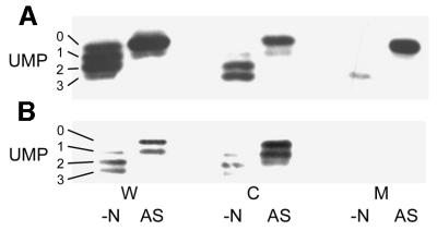
Fig. 3. The membrane association of GlnK reflects the uridylylation state of the protein. Whole-cell extracts (W), and cytoplasmic (C) and membrane (M) fractions were prepared from strains (A) ΔglnB (PT8000) and (B) ΔglnB,ΔamtB (AT8000) both before (–N) and after (AS) ammonia shock. Extracts and fractions were prepared as described in Materials and methods and subjected to native PAGE followed by western blotting using an anti-PII antibody. Under these conditions, all four possible forms of GlnK (UMP 0–3) can be identified.

An official website of the United States government
Here's how you know
Official websites use .gov
A
.gov website belongs to an official
government organization in the United States.
Secure .gov websites use HTTPS
A lock (
) or https:// means you've safely
connected to the .gov website. Share sensitive
information only on official, secure websites.
