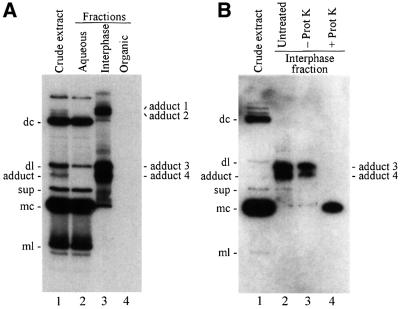
Fig. 3. Northern blot analysis of extracts from ASBVd-infected UV-irradiated avocado leaves. Extracts were fractionated by denaturing PAGE and hybridized with a probe for detecting (+) viroid strands. (A) Partition after treatment with phenol/chloroform. Lane 1, crude extract; lanes 2, 3 and 4, fractions corresponding to the aqueous phase, interphase and organic phase, respectively. The interphase and organic phase fractions were concentrated 20-fold by alcohol precipitation with respect to the crude extract and the aqueous phase fraction. (B) Proteinase K digestion of a preparation enriched in adducts 3 and 4 derived from the concentrated interphase fraction. Lane 1, control of crude extract; lanes 2, 3 and 4, preparation untreated, incubated in buffer without proteinase K, and digested with proteinase K, respectively. The preparation enriched in adducts 3 and 4 was obtained by cutting a segment of a non-denaturing polyacrylamide gel and eluting its contents. The positions of the different ASBVd (+) RNAs and of the four ASBVd–protein cross-linked adducts are indicated on the left and right sides of both panels. Other details as in the legend to Figure 1.

An official website of the United States government
Here's how you know
Official websites use .gov
A
.gov website belongs to an official
government organization in the United States.
Secure .gov websites use HTTPS
A lock (
) or https:// means you've safely
connected to the .gov website. Share sensitive
information only on official, secure websites.
