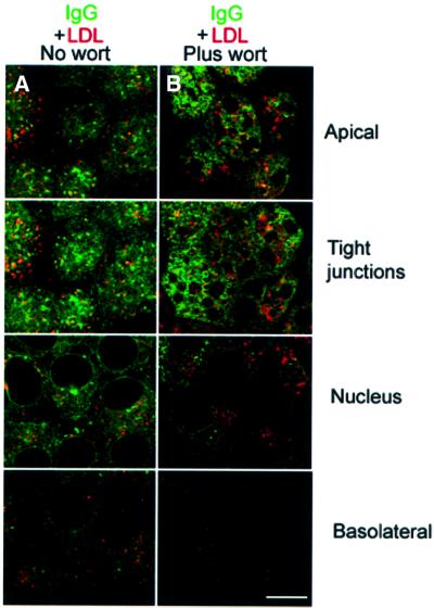
Fig. 7. Co-localization of FcRn–GFP and LDL. Bar, 5 µm. Sections labeled as apical, tight junctions, nucleus and basolateral were taken 1, 3, 6 and 9 µm, respectively, from the apical surface of the monolayer. Cells were serum starved and incubated at 37°C with Alexa 646 IgG and DiI-LDL at the apical surface for 30 min in the absence (A) and presence (B) of 150 nM wortmannin. Regions of co-localization appear yellow.

An official website of the United States government
Here's how you know
Official websites use .gov
A
.gov website belongs to an official
government organization in the United States.
Secure .gov websites use HTTPS
A lock (
) or https:// means you've safely
connected to the .gov website. Share sensitive
information only on official, secure websites.
