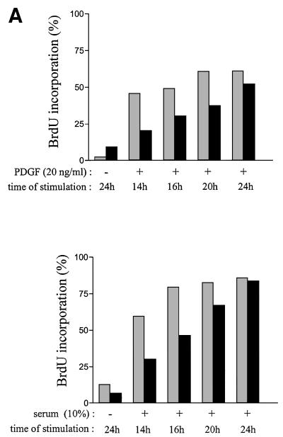
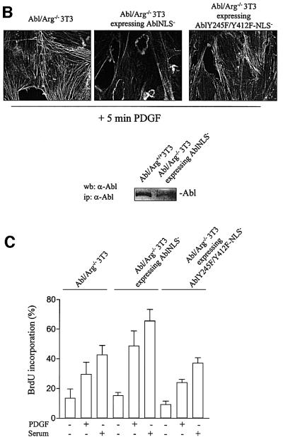
Fig. 6. Quiescent Abl/Arg–/–3T3 fibroblasts exhibit a 4–6 h delay in S phase entry when induced by PDGF or serum. (A) Cells deficient in Abl and Arg exhibit a 4–6 h delay in S phase entry induced by mitogens. Abl/Arg+/+ 3T3 (grey bars) and Abl/Arg–/– 3T3 (black bars) cells were grown on coverslips and made quiescent by serum starvation. Cells were stimulated or not with PDGF (top panel) or serum (bottom panel), as indicated in the presence of BrdU for indicated times, fixed, stained for BrdU incorporation by immunostaining and analysed by microscopy. Shown is the percentage of BrdU-positive cells under the specified conditions, calculated as in Figure 2. These data are representative of two independent experiments. (B) Abl-NLS– restores the actin reorganization induced by PDGF in Abl/Arg–/– 3T3 cells. Top panel: Abl/Arg–/– 3T3 and Abl/Arg–/– 3T3 expressing the indicated constructs grown onto coverslips were made quiescent by serum starvation, stimulated with PDGF for 5 min and fixed. Actin was visualized by staining with rhodamin–phalloidin. Pictures are representative of two independent experiments. Bottom panel: level of immunoprecipitated Abl from the cytoplasmic fraction of the indicated cells. (C) Abl-NLS– restores mitogenesis in Abl/Arg–/– 3T3 cells. Cells were stimulated or not with PDGF (20 ng/ml) or serum (10%), as indicated, in the presence of BrdU for 16 h, then fixed and stained for BrdU incorporation and analysed by microscopy. Shown is the percentage of BrdU-positive cells under the specified conditions, calculated as described in Figure 2. The data from three independent experiments have been averaged, and the mean and standard deviation are shown.

An official website of the United States government
Here's how you know
Official websites use .gov
A
.gov website belongs to an official
government organization in the United States.
Secure .gov websites use HTTPS
A lock (
) or https:// means you've safely
connected to the .gov website. Share sensitive
information only on official, secure websites.

