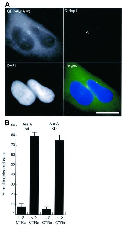
Fig. 3. Aurora-A overexpression causes multinucleation concomitant with centrosome amplification. (A) HeLa cells were transfected for 48 h with wt or KD mutant EGFP–Aurora-A and analyzed by immunofluorescence microscopy. Transfected cells were identified by GFP-fluorescence (green), centrosomes were stained with anti-C-Nap1 antibodies (red) and DNA with DAPI (blue). Scale bar: 10 µm. (B) Transfected cells were classified according to whether they had normal numbers of centrosomes (one or two fluorescent dots) or more than two centrosomes, and whether they were mononucleated or multinucleated. Histogram shows results from three independent experiments (400–600 cells each) and bars indicate standard deviations.

An official website of the United States government
Here's how you know
Official websites use .gov
A
.gov website belongs to an official
government organization in the United States.
Secure .gov websites use HTTPS
A lock (
) or https:// means you've safely
connected to the .gov website. Share sensitive
information only on official, secure websites.
