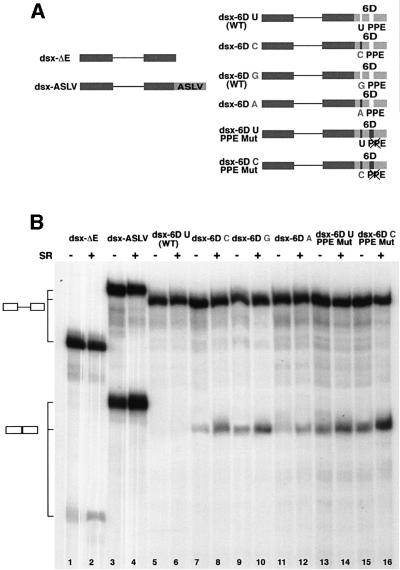
Fig. 3. In vitro splicing of the dsx–exon 6D substrates. (A) Schematic representation of the dsx–exon 6D splicing substrates. Dsx exonic sequences: dark boxes; ASLV and exon 6D sequences: light boxes. U-to-C, U-to-G, U-to-A and PPE mutations are indicated. (B) In vitro splicing reactions. The radiolabeled pre-RNA substrates indicated were spliced in in vitro splicing reaction mixtures containing HeLa cell nuclear extract. Purified HeLa cell SR proteins (200 ng) were added where indicated. After 2 h of incubation at 30°C, the RNAs were recovered and separated on a 6% polyacrylamide–8 M urea gel. RNA precursors and spliced products are indicated by the schematics on the left.

An official website of the United States government
Here's how you know
Official websites use .gov
A
.gov website belongs to an official
government organization in the United States.
Secure .gov websites use HTTPS
A lock (
) or https:// means you've safely
connected to the .gov website. Share sensitive
information only on official, secure websites.
