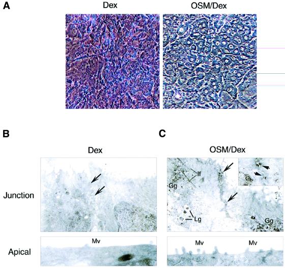
Fig. 1. Morphological changes induced by OSM. Fetal hepatocytes derived from murine liver at E14.5 were cultured for 6 days with Dex or OSM/Dex. (A) Phase contrast images of cultured hepatocytes. OSM/Dex-stimulated cells exhibited tight cell–cell contact, a dense cytoplasm and a clear and round nucleus. (B and C) Ultrastructure of fetal hepatocytes. Cells cultured with Dex (B) or OSM/Dex (C) were processed for electron microscopy. Upper panels: junction areas between adjacent cells. Arrows indicate junctional zones between adjacent cells. Markers shown are Lg, lipid granule; Gg, glycogen granule; If, intermediate filament. The insert in (C) shows desmosome-like structures that are indicated by arrows. Lower panels: apical membrane domain. Mv, microvilli.

An official website of the United States government
Here's how you know
Official websites use .gov
A
.gov website belongs to an official
government organization in the United States.
Secure .gov websites use HTTPS
A lock (
) or https:// means you've safely
connected to the .gov website. Share sensitive
information only on official, secure websites.
