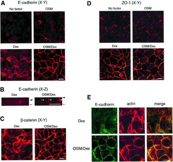
Fig. 2. OSM induces formation of adherens junctions, but not tight junctions. Fetal hepatocytes were cultured for 6 days without OSM/Dex (No factor) or with OSM, Dex or OSM/Dex. The localization of cell adhesion molecules was examined by immunofluorescence analysis. (A and B) Subcellular distribution of E-cadherin in cultured hepatocytes. Cells were fixed and stained with anti-E-cadherin antibody. (A) x–y views; (B) x–z views of the cells. Bars and arrows indicate the bottom and top of the cells, respectively. E-cadherin was highly concentrated at the apical parts of lateral membrane (arrowheads). (C and D) Intracellular localization of β-catenin and ZO-1. Cells were fixed and stained with antibodies against β-catenin (C) or ZO-1 (D). (E) Co-localization of E-cadherin and the actin cytoskeleton. Cells were stained with anti-E-cadherin antibody (green, left panels) and rhodamine–phalloidin (red, middle panels). Each image was overlaid (yellow, right panels). Upper panels: Dex-treated cells. Lower panels: OSM/Dex-treated cells. Scale bars: 10 µm.

An official website of the United States government
Here's how you know
Official websites use .gov
A
.gov website belongs to an official
government organization in the United States.
Secure .gov websites use HTTPS
A lock (
) or https:// means you've safely
connected to the .gov website. Share sensitive
information only on official, secure websites.
