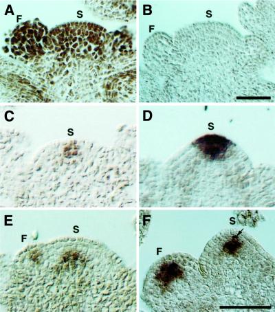
Fig. 7. Expression pattern of SHD, CLV3 and WUS mRNA in the SAM and FM. (A) Wild-type (Ler) SAM and FM hybridized with an antisense SHD probe. SHD mRNA is distributed throughout the SAM and FM. (B) Wild-type SAM and FM hybridized with a sense SHD probe. (C) Wild-type SAM hybridized with an antisense CLV3 probe. CLV3 mRNA is localized in a few cells at the SAM. (D) shd SAM hybridized with an antisense CLV3 probe. The CLV3 expression domain is enlarged compared with the wild type (C). (E) Wild-type SAM and FM hybridized with an antisense WUS probe. WUS mRNA is detected in a small group of cells both in the SAM and FM. In the SAM, the WUS expressing cells localize underneath the three outermost cell layers. (F) shd SAM and FM hybridized with an antisense WUS probe. The WUS expression domains are enlarged both in the SAM and FM, and WUS mRNA is detected even in the third layer of the SAM, i.e. one cell layer up compared with the wild type (E). In this panel, a second-layer cell (arrow) in the SAM is also stained. (A) and (B) are the same magnifications, and (C–F) are the same magnifications. Bar: 50 µm. S, SAM; F, FM.

An official website of the United States government
Here's how you know
Official websites use .gov
A
.gov website belongs to an official
government organization in the United States.
Secure .gov websites use HTTPS
A lock (
) or https:// means you've safely
connected to the .gov website. Share sensitive
information only on official, secure websites.
