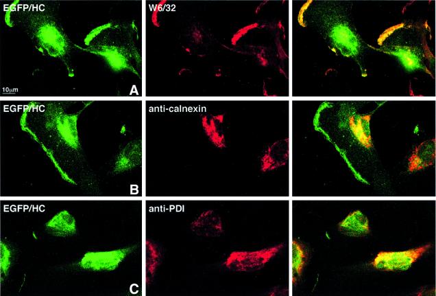
Fig. 2. Intracellular distribution of EGFP–HC is similar to that of endogenous class I molecules. U373EGFP–HC were grown on chamber slides over night, prior to fixation, immunohistochemistry and confocal laser scanning microscopy. Double-positivity of the green fluorescent EGFP–HC molecule with W6/32 corresponds to properly folded class I complexes that contain the chimeric molecule. These are found at the cell surface (A) and in the ER. ER localization of EGFP–HC is demonstrated by staining with the ER markers calnexin (mAb AF8) (B) and PDI (anti-PDI) (C). For each antibody the green EGFP–HC (left column), the respective second staining in red (middle column) and a merge image (right column) are shown, as indicated for the individual panels.

An official website of the United States government
Here's how you know
Official websites use .gov
A
.gov website belongs to an official
government organization in the United States.
Secure .gov websites use HTTPS
A lock (
) or https:// means you've safely
connected to the .gov website. Share sensitive
information only on official, secure websites.
