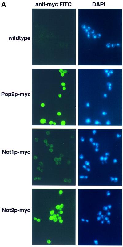
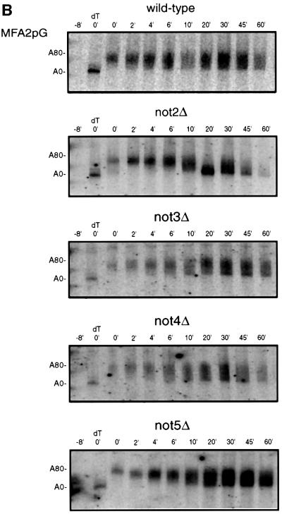
Fig. 7. The Not protein complex functions in deadenylation. (A) Localization of Not1p and Not2p by indirect immunofluorescence. Strains expressing myc-tagged Not1p (yRP1668), myc-tagged Not2p (yRP1669), myc-tagged Pop2p (yRP1622), or wild-type control (yRP841), were stained with anti-myc antibodies and anti-mouse (FITC) secondary antibody and DAPI as indicated. It is critical to note that all photographs were taken under identical conditions. The localization of myc-tagged Not1p was consistently cytoplasmic; however, the signal often appeared weaker. This observation is presumably a result of the Not1p myc-epitope being less accessible to the anti-myc antibodies. (B) Transcriptional pulse–chase analysis of the MFA2pG in not2Δ, not3Δ, not4Δ and not5Δ strains. Shown are polyacrylamide northern gels examining the decay of MFA2pG in the indicated strains. Numbers above the lanes are minutes after transcriptional repression by the addition of glucose following an 8 min induction of transcription (Decker and Parker, 1993). The 0 min time points were treated with RNase H and oligo(dT) to indicate the position of the deadenylated mRNA.

An official website of the United States government
Here's how you know
Official websites use .gov
A
.gov website belongs to an official
government organization in the United States.
Secure .gov websites use HTTPS
A lock (
) or https:// means you've safely
connected to the .gov website. Share sensitive
information only on official, secure websites.

