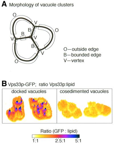
Fig. 4. Vertex enrichment of selected proteins during docking. (A) Membrane microdomains of docked vacuoles are defined as ‘outside edges’, which are not in contact with other vacuoles, ‘boundary edges’, which are opposed to other vacuoles in the cluster, and ‘vertices’, where boundary edge and outside edge membranes meet. (B) Docking-dependent enrichment of Vps33p at vertices. Left panel: purified vacuoles from cells with GFP-tagged Vps33p were stained with FM4-64, incubated in a standard fusion reaction for 30 min to allow docking and examined by ratiometric fluorescence microscopy to determine regions of membrane where tagged Vps33p is enriched. Right panel: vacuoles were not incubated under docking and fusion conditons with ATP, but rather were clustered by co-sedimentation. Reprinted with permission from Elsevier Science from L.Wang et al. (2002).

An official website of the United States government
Here's how you know
Official websites use .gov
A
.gov website belongs to an official
government organization in the United States.
Secure .gov websites use HTTPS
A lock (
) or https:// means you've safely
connected to the .gov website. Share sensitive
information only on official, secure websites.
